Welcome to Euro-BioImaging Access Portal
Get access to imaging technologies and image data analysis across Europe - open to everyone!
Applying for access is simple and quick. After you log in, submit a short proposal. Click here to learn more.
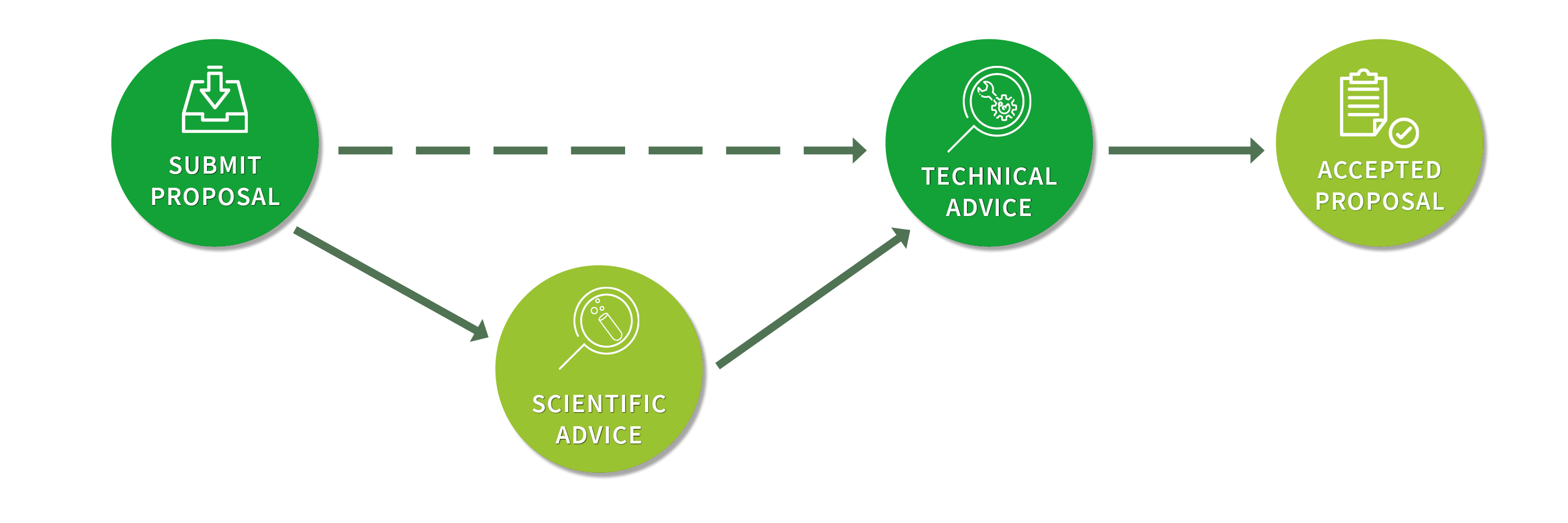
You can also ask us for advise on what type of imaging suits you and where to find it.