NETHERLANDS
Advanced NL Microscopy Node
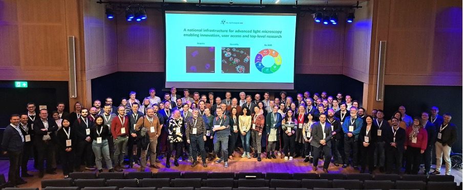
The Advanced NL Microscopy Node is a multimodal, multi sited Euro BioImaging Node that unites leading Dutch centres for advanced light and electron microscopy and multi modal molecular imaging. It consists of 10 advanced microscopy facilities distributed across the Netherlands and is coordinated by Amsterdam UMC as the Representing Entity.
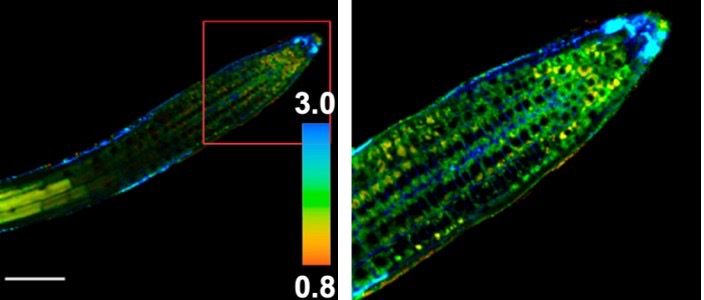
Application focus areas span basic and translational biomedical research (cell biology, cancer, neuroscience, cardiovascular, immunology) as well as plant and agricultural imaging, where Wageningen brings unique molecular and functional imaging expertise. Distinctive strengths include national‑scale correlative microscope (CLEM) workflows, high‑content screening for functional genomics and drug discovery, optogenetic probe and self‑steering microscopy development, and advanced functional imaging combined with custom biosensor and fluorophore design.
Strategically, the Node was created by merging five previous Dutch Euro‑BioImaging Nodes (CLEM, LCAM: the Amsterdam Leeuwenhoek Center for Advanced Microscopy, High Throughput Microscopy, WISH: Wageningen Imaging and Spectroscopy Hub, Erasmus MC Optical Imaging Centre) into one unified Advanced NL Microscopy Node. This merged Node provides a single, coordinated entry point for Euro‑BioImaging users, unified access policies, shared data‑management and analysis pipelines, and an integrated training programme.
All facilities are part of NL-BioImaging (NL-BI) which is a Dutch distributed research infrastructure aimed at promoting progress in the most cutting-edge light microscopy technologies and providing open access to these technologies to academic and industrial users.
Specialties and expertise of the Node
The Advanced NL Microscopy Node unites 10 collaborating facilities:
1. Combined correlative microscopy using light and electron microscopy (CLEM)
- Cell Microscopy Center UMC Utrecht
- LUMC Electron microscopy lab
- UMC Groningen Microscopy and Imaging Center (UMIC)
2. Application of F-techniques (FCS, FLIM, FRAP, FRET) and the development of optogenetic probes, self-steering microscopy and
- Erasmus MC OIC
- LCAM-AmsterdamUMC / MCCF AmsterdamUMC–AMC
- LCAM-FNWI Amsterdam
- LCAM-NKI Amsterdam
- Wageningen Imaging and Spectroscopy Hub (WISH)
3. High-content microscopy
- Leiden Cell Observatory
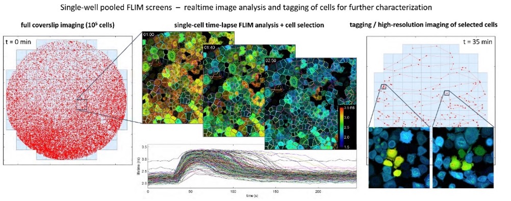
- NKI Robotics and Screening Center Amsterdam
4. Functional imaging technologies and their applications in plant/agricultural imaging
- Wageningen Imaging and Spectroscopy Hub (WISH)
Offered Technologies
Advanced light microscopy
- Laser scanning confocal microscopy (LSCM/CLSM)
- Spinning disk confocal microscopy (SDCM)
- Structured illumination microscopy (SIM)
- Total internal reflection fluorescence microscopy (TIRF)
- Two-photon microscopy (2P)
- Single Molecule localisation microscopy (SMLM)
- Light-Sheet Microscopy
- Stimulated Emission by Depletion (3D-STED)
- High throughput microscopy/high content screening (HTM/HCS) confocal and widefield
- Multiplex imaging (confocal and widefield)
- Laser microsdissection (LMD)
- Confocal Raman Microscopy (CRM)
- Visual omics / spatial omics
F-techniques
- Fluorescence Resonance Energy Transfer (FRET)
- Fluorescence Recovery After Photobleaching (FRAP)
- Fluorescence (cross)-correlation spectroscopy (FCS/FCCS)
- Fluorescence Lifetime Imaging Microscopy (FLIM)
Transmission and scanning electronmicroscopy
- TEM of chemical fixed and cryo-immobilized (HPF)samples
- EM tomography
- serial section TEM and blockface SEM
- FIB-SEM
- STEM and SEM tomography
- Elemental analysis
- Array tomography
- Immuno-gold EM on resin and thawed cryo-sections (Tokuyasu Method)
- Genetic encoded EM probes
- Pre-embedding immunolabelling
- Cryo TEM and ET
Correlative light and electron microscopy (CLEM)
- pre-embed and on-section CLEM
- Cryo-CLEM
- live-cell CLEM
- EDX-CLEM (Correlative EDX and SEM)
Data management and analysis
- Smart microscopy scripts
- FAIR data management
- Post-acquiring data analysis
Additional Services offered by the Node
- Assistance with experimental design (e.g. experimental set up, choice of technique)
- Execution of pilot studies by node staff
- Training in sample preparation
- Training in use of light and electron microscopes
- Assistance of users in the execution of experiments
- Wet lab space
- Data processing and analysis software
- Desk space and training seminar room
- Cell culture facilities
- Technical assistance to run instrument
- Training in infrastructure use
- Probe preparation
- Animal preparation
- Data processing and analysis
- Training workstations
- Biobanking, biological material storage and processing
- Cell culture facilities - Safety level 1 and 2
- Assistance with Crispr libraries
- Assistance with assay development
Instrument highlights
At Cell Microscopy Center UMC Utrecht:
At Koster Lab - LUMC Electron microscopy research
At LCAM-AmsterdamUMC / MCCF AmsterdamUMC–AMC
· Confocal: Leica SP8 X WLL, Leica SP8-SMD (Live-Cells, FLIM, FRAP) WLL, Leica SP8-DLS (LightSheet) WLL, Leica SP5-II (multibeam), Leica Spectraplex
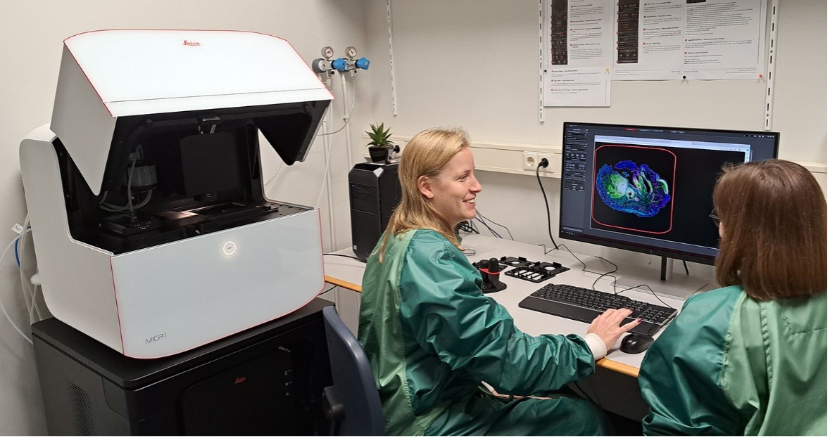
· Widefield: Leica MICA, Leica DMi8(Live Cells), Leica Thunder, Leica DM6, Leica IRBE (Microinjection), Leica Cell DIVE multiplex, Molecular Devices ImageXpress Pico (High-Content Imager, Zeiss Axio Zoom
· Leica LMD7 laser micro dissection
· Electron Microscopy: Talos L120c with EDX, Tecnai G2 Spirit Biotwin, JEM-1400 Flash, Zeiss Gemini Sigma 300 (Scanning)
· Flow Cytometry: analysers BD FACSCanto II (3 lasers), BD Symphony A1 (4 lasers), Sony ID7000 Spectral Cytometer (6 lasers), Cytek Amnis ImageStream X Mk II; sorters Sony FP7000 spectral sorter (6 lasers), Sony SH800 Cell Sorter (4 laser), BD Symphony S6 (4 laser)
At LCAM-FNWI Amsterdam
· Confocal: Leica SP8, Zeiss LSMmeta/Confocor, Zeiss LSM510, Nikon A1 (CLEM, FRAP), Olympus FLIM (FLCCS) Spinning Disk Confocal: Zeiss Yokogawa (FLIM), Nikon Andor (TIRF)
· Widefield: Zeiss Cell Observer, Nikon/Lambert Instruments FLIM screener, Zeiss/Lambert Instruments FLIM/SPIM
· Superresolution: Nikon Andor-PALM
At LCAM-NKI Amsterdam
At Leiden Cell Observatory
· 2 x LIPSI NIKON systems, 1 x NIKON C2 systems,1 x NIKON A1 system equipped with FLIM, 1 x Zeiss Cell Observer, 1 X ImageXpress.ai system.
At Microspectroscopy Research Facility Wageningen (WISH)
· Confocal: Leica SP8 SMD, Leica SP8, Leica SP5 (upright) Leica Stellaris 5, Leica Stellaris-STED, Leica MP-Dive-FLIM, Nikon C2, RCM confocal, Zeiss 710,Zeiss 900, Piqoquant confocal-optical tweezer, Roper Spinning Disk Confocal,
· Widefield: Nikon, Homebuilt TIRF Andor-PALM, Raman microscope
At NKI Robotics and Screening Center Amsterdam
· Stellaris FALCON/STED instrument with 355nm and 405 nm external lasers for fast UV photolysis/photoactivation experiments
At Optical Imaging Centre Rotterdam
· Confocal: Zeiss Meta 729, LSM 700 and 900, Leica SP5 and SP8, Opera Phenix HCS system
· Widefield: Nikon TIRF 1454 and Spinning Disc 1454, Zeiss Elyra PS1 SIM, InVi SPIM lightsheet
At UMC Groningen Microscopy and Imaging Center (UMIC) TEMs, SEMs, STEM and EDX detectors, FIB-SEMs, (live-cell) FMs, High-pressure freezers (HPFs), Vitrification equipment, Automated Freeze Substitution Devices, Ultramicromomes, Cryo-ultramicrotomes.
Contact details
Eric Reits
Advanced NL Microscopy Node Management
List of sites
Facility | Former Node | Contact | |
1. Cell Microscopy Center UMC Utrecht | CLEM | Nalan Liv | |
2. Koster Lab - LUMC Electron microscopy research | CLEM | Roman Koning | |
3. LCAM-AmsterdamUMC / MCCF AmsterdamUMC–AMC | LCAM | Eric Reits | |
4. LCAM-FNWI Amsterdam | LCAM | Mark Hink | |
5. LCAM-NKI Amsterdam | LCAM | Bram van den Broek |
|
6. Leiden Cell Observatory | HTM | Sylvia Le Devedec | |
7. Microspectroscopy Research Facility Wageningen | WISH | Jan Willem Borst | |
8. NKI Robotics and Screening Center Amsterdam | HTM | Roderick L. Beijersbergen | |
9. Optical Imaging Centre Rotterdam | OIC | Gert-Jan Kremers | |
10. UMC Groningen Microscopy and Imaging Center (UMIC) | CLEM | Ben Giepmans |