Laser Speckle Contrast Imaging
Laser Speckle Contrast Imaging – also known as Laser Speckle Contrast Analysis – is a non-invasive optical technique combining high resolution with high speed. This provides real-time determination of local microcirculation in the whole examination area, which can be performed in parallel with the dynamic analysis of the vascular response.
Laser Speckle Contrast imaging is based on a coherent laser light which, when hitting a diffuse object having a surface with imperfections, produces a random interference pattern known as speckle pattern, which is monitored by a CCD camera. If the scattering medium is static, the speckle pattern remains stable over time. However, if there is motion – for example circulating blood cells – the speckle pattern fluctuates in intensity leading to a reduction in the observed speckle contrast and providing real-time information about the movement.
A key advantage of this imaging technique is that it provides full-field, real-time mapping of relative flows without the need of exogenous contrast agents.
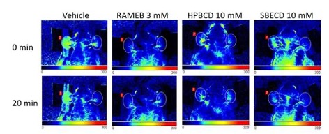
Therefore, this technique is highly suitable for the evaluation of blood perfusion via the analysis of speckle pattern variations and representation as real-time graphs as well as color-coded images.
In preclinical research, Laser Speckle Imaging can be widely used for investigating dermal perfusion, skin inflammation and wound healing, as well as ischemic and neuropathic states of the extremities and acute and chronic inflammatory disorders of the joints. Additional application areas include cerebral blood flow monitoring, as well as models of stroke and migraine.
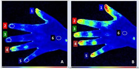
In clinical research and practice, this technique provides good opportunities for the assessment of dermatological disorders (e.g. allergic skin reactions), endothelial dysfunction of various origins, skin cancers, vasospastic conditions such as Raynaud’s phenomenon, as well as peripheral arterial disease and its more severe form, chronic limb threatening ischemia.
doi: 10.1016/j.jlr.2025.100844
doi: 10.3325/cmj.2017.58.424.
Laser Speckle Contrast Imaging is provided by the Medical and Preclinical Imaging Hungary Node and by Danish BioImaging.
Use cases | Node | DOI |
Skin investigation in animal models: acute skin inflammation | ||
Investigation of joint inflammation in animal models | ||
Investigation of the hypothermic effect of H2S in mice | ||
Investigation of a dermatitis model of psoriasis | https://doi.org/10.1002/cpph.78 | |
Investigation of a clinical case of cold agglutinin-induced acrocyanosis | ||
Microvascular alterations in human nonlesional psoriatic skin |